


Strengthen and stabilise a weakened cornea with C3R (Corneal Collagen Cross-Linking with Riboflavin), a treatment used to help slow the progression of keratoconus.
Get your Booking to get emergency service
C3R, also known as corneal collagen cross-linking (CXL), is a treatment used to strengthen the cornea in patients with keratoconus. Keratoconus causes the cornea to become thinner and gradually change into a more cone-like shape, which can lead to blurred or distorted vision.
During C3R treatment, riboflavin (vitamin B2) eye drops are applied to the cornea and activated using controlled ultraviolet-A light. This process increases the bonding between collagen fibres within the cornea, helping strengthen its structure and slow further progression of keratoconus.

C3R helps strengthen the weakened corneal structure and can help slow the progression of keratoconus.

By increasing collagen cross-linking within the cornea, the treatment helps maintain greater structural stability.

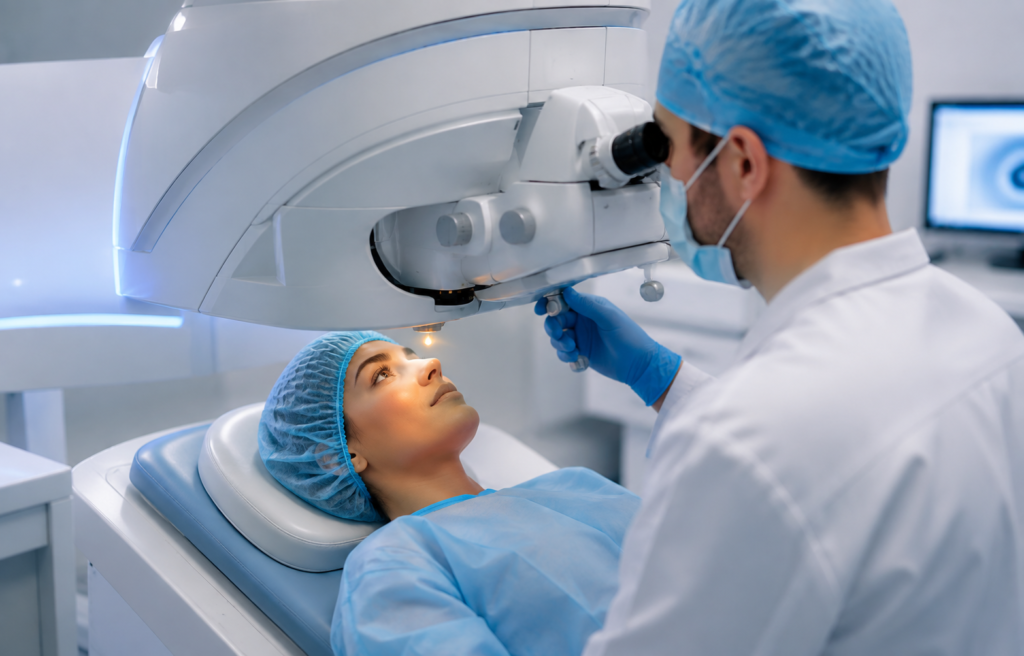
C3R is performed using riboflavin eye drops and controlled ultraviolet-A light without removing corneal tissue.

Early treatment may help prevent further corneal weakening and reduce the risk of progressive visual deterioration.

C3R works by strengthening the existing corneal collagen rather than replacing or removing the cornea.

A detailed corneal examination and mapping help the ophthalmologist assess the condition and determine the appropriate treatment approach.

SC3R is generally considered for patients with keratoconus where examination shows evidence of progression or a risk of further corneal weakening.
C3R may be recommended when tests show that keratoconus is progressing and the cornea is becoming weaker or more irregular.
Patients with a structurally weakened cornea may be assessed for cross-linking to help strengthen and stabilise the corneal tissue.
A comprehensive eye examination, corneal topography and assessment of corneal thickness help determine whether C3R is appropriate for the individual patient.
Every patient undergoes a detailed corneal evaluation to determine whether C3R
is the most appropriate treatment option.
Anaesthetic Eye Drops
The eye is numbed with local anaesthetic drops for comfort during treatment.
Corneal Preparation
The cornea is prepared according to the treatment protocol recommended by the ophthalmologist.
Riboflavin Application
Riboflavin (vitamin B2) eye drops are applied to the cornea to allow adequate absorption.
UV-A Light Exposure
Controlled ultraviolet-A light is applied to activate the riboflavin and promote collagen cross-linking.
Corneal Stabilisation
The cross-linking process strengthens the corneal structure and helps slow the progression of keratoconus.
C3R may be suitable for patients with keratoconus where clinical examination and corneal imaging indicate progression or a need for corneal strengthening. Treatment suitability depends on factors such as corneal thickness, corneal shape, disease progression and overall eye health.
A detailed corneal examination is essential before treatment. Your ophthalmologist will review your corneal mapping and other test results to determine whether C3R is appropriate for your eyes.
At Dr. P. S. Hardia Advanced Eye Surgery and Research Institute, patients with keratoconus undergo detailed corneal evaluation before treatment to support accurate diagnosis and personalised treatment planning.
Detailed corneal mapping helps assess corneal shape, thickness and changes associated with keratoconus.
Cornea and refractive specialists assess each patient's condition and recommend treatment according to individual clinical findings.
Detailed examination and follow-up help monitor corneal stability and assess treatment response over time.
Improve your vision and stabilize corneal shape with precision-engineered INTACS the advanced surgical alternative to corneal transplants.
Get your Booking to get emergency service
At Dr. P. S. Hardia Advanced Eye Surgery and Research Institute, we offer INTACS (Intracorneal Ring Segments), a breakthrough technology specifically designed for the management of Keratoconus. Keratoconus is a progressive condition where the cornea thins and bulges into a cone-like shape, causing severe visual distortion. INTACS are microscopic, clear segments inserted into the corneal stroma to flatten the bulge and restore a more natural, spherical shape to the eye.
This procedure is ideal for patients who are intolerant to contact lenses or for those whose Keratoconus is progressing despite other treatments. Unlike traditional surgeries, INTACS do not involve removing any corneal tissue; instead, they act as structural supports to reinforce the cornea. This effectively reduces irregular astigmatism and myopia, often significantly improving the patient’s best-corrected vision and quality of life.
Backed by our Guinness World Record legacy and extensive experience in complex corneal cases, we provide a personalized approach to INTACS implantation. We often combine INTACS with C3R (Corneal Collagen Cross-linking) for a dual-action result: C3R to stop the progression and INTACS to improve the visual shape. Our commitment to using high-definition corneal mapping ensures that every segment is placed with microscopic precision for maximum visual stability.
By utilizing ultra-thin, biocompatible ring segments and femtosecond laser precision, we reshape the irregular cornea from within, providing a stable structural foundation that enhances visual clarity and contact lens comfort.
About Us
At Dr. P. S. Hardia Advanced Eye Surgery and Research Cente, we are dedicated to delivering exceptional healthcare with compassion, innovation, and integrity. For over 20 years, we have been a offering comprehensive medical services
Contact Info